Lesson 10
Miscellaneous Diseases of the Eye and other
Special Anatomical Areas
Diseases of the Eye
I. Entropion (turning inward of the eyelid,
usually lower lid)
A.
Normally seen in newborn foals.
B.
May correct itself, but if eye irritation is severe or corneal ulcer develops,
surgical correction is necessary.
II. Eyelid trauma (tearing of eyelid)
A.
Surgical repair should be preformed promptly if possible.
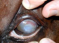
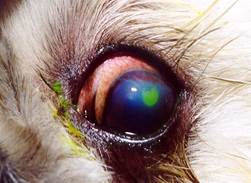
III. Corneal Ulceration
A.
A fairly common injury in the horse.
B.
Requires veterinary attention
C.
Signs of corneal ulceration include:
1.
Excessive tearing.
2.
Evidence of pain of the eye in the way of squinting and closing the lid.
3.
Cloudy or bluish tint to the eyeball.
D.
May be caused by a bacterial infection or fungus. The white blood cells that
are produced enhance the progression of the ulcer. This action is called “melting”
and is apparent by the gray liquid that forms around the ulcer. This is an emergency situation and a
veterinarian must be called immediately.
E.
Treatment involves continuous application of antibiotic ointment and atropine
ointment twice a day.
F.
Unless horse is not adequately cared for when dealing with a corneal ulcer, the
horse may loose the eye.
G.
Corticosteroid ointments (cortisone) are not to be used in corneal ulcers, and
only by veterinarian recommendation for other eye conditions.
IV. Conjunctivitis
A.
Normally caused by fly irritation and secondary bacterial infection.
B.
Antibacterial ointments and occasionally antibacterial with corticosteroid
ointments recommended.
C.
Fly control important as prevention.
V. Equine Recurrent Uveitis
(ERU) (Moon
Blindness)
Appaloosas are eight times more
likely to contract recurrent uveitis.
Possible genetic link, currently being researched
Three
types of ERU: classic, insidious, and posterior.
A.
Classic: most common type. Symptoms are pain and inflammation, followed by an
inactive period of time which has no limit on length of time, then a reoccurrence
of the symptoms. This cycle will
continue until the horse goes blind.
B.
Insidious: generally no signs of pain. A
low-grade inflammation remains, even during the quiet cycle, which continues to
damage the eye. By the time the owner
decides to call the vet, the eye may be too far damaged to save.
C.
Posterior: not often seen in the United States.
Inflammation develops behind the lens. Eventually the retina will detach.
Causes:
research is on-going, possibly genetic.
Injury, environment, illness/infection may trigger the disease.
Treatment:
reduce inflammation and prevent ocular damage. Use of NSAISs
and corticosteroids. Start treatment early and aggressively. Wean horses off drugs as soon as possible to
prevent side effects (digestive ulcers).
Implantation of a device that releases medication at a regulated dose
may be an option. Even with aggressive
treatment horses will go blind eventually.
Management:
protect eyes from sunlight, limit exposure to dust, avoid using hay nets or
raised hay feeders, and remove any objects that could damage the eye (low
branches, protruding nails, bucket hooks, splintered boards), protect eyes
during transporting, control flies.
Consider
the following symptoms cause for alarm and call the veterinarian.
1.
A red swollen eye
2.
Excessive squinting
3.
Excessive tearing
4.
Increased sensitivity to bright light
5.
Cloudy or blue appearance to cornea.
Physical Problems of the Spine and
Back
I. Fractures of the Spine
A. The dorsal thoracic vertebrae, with their tall
dorsal projections, can be associated with fractures during falls. There is
usually readily identifiable pain and inflammation in the area. X-rays usually indicate the amount of damage. Healing can take up to 6 months.
B.
Fractures, which would involve the spinal chord, would obviously cause a grave
prognosis due to paralysis problems.
II. Cervical Vertebral
Malformation (Wobbles, Bobbie)
A.
Can be associated with OCD (developmental disease of young, growing horses).
B.
Causes pressure on the cervical spinal chord, causing partial paralysis,
in-coordination and weakness in the hind quarters. (They can’t seem to control their
hindquarters, especially while turning or stopping.)
C.
Usually a permanent and progressive problem, the horse may ultimately be found
down and unable to get up.
D.
Treatment is usually unsuccessful, unless a very complicated surgery is
attempted.
III.
Scoliosis
–Lateral to medial deviation, versus Lordosis – Sway backed. Both are
congenital (horse is born with the problem).
IV. Hunter’s Bumps
A.
Lumbar and Sacral dorsal prominences and their respective ligamentous
attachments are involved in hunter’s bumps. They usually appear at the highest
point of the horse’s croup. Strain in
this area causes atrophy, tearing and subluxation of muscle attachments, which
produces the characteristic hunter’s bump. Hind limb lameness is also apparent.
Pain
is experienced by the horse during the acute stage. Stability to the joint will
resume once the torn ligaments form scar tissue. The hunter’s bump is evidence of a previous
injury.
Seen in hunters and jumpers
predominantly.
V. Sacroiliac subluxation
A.
A strain of the left or right side of the sacroiliac joint area, which will
cause a raising of one side or the other of the pelvic
(illium) bone.
Also seen in horses used in jumping events. When it first occurs there will be associated
swelling and pain in the area. Can take up to
Muscle Conditions of the Horse
I. Muscle Atrophy
A.
Lack of nerve supply to the muscle.
B.
Lack of blood supply to the muscle.
C.
Lack of use of the muscle.
D.
Trauma or damage to the muscle.
E.
Severe overuse of the muscle.
II. Myositis (sore back)
A.
Inflammation of the muscle tissue. (Overuse, overstretching, trauma to the muscle).
B.
Usually associated with the back, loin and croup areas of the horse.
C.
Most often associated with a chronic situation which needs to be managed.
D.
NSAID’s, heat therapy, liniment therapy, massage therapy, muscle relaxants,
chiropractic manipulations, and other forms of therapy are in common use.
III. Rhabdomyolysis (tying up)
Clinical signs – varying
degrees of soreness from just being stiff to intense pain rendering the horse
incapable of moving, standing or bearing weight. The most common area affected is the hindquarters.
Hard painful muscles and cramping apparent. Profuse sweating; elevated heart and respiratory rates. Colic-like behavior. Severe cases: dark, red-brown colored
urine.
Prior
to more research tying up was considered to be one disease, commonly called
Monday Morning disease (draft horses were prone to tying up after a weekend of
rest) or azoturia.
It is now put in two categories: Sporadic Exertional Rhabdomyolysis and
Chronic Exertional Rhabdomyolysis.
A.
Sporadic Exertional Rhabdomyolysis
1.
Causes: exercise beyond the horse’s fitness level; nutrient deficiencies of
sodium, Vitamin E and selenium; inverted calcium to phosphorus ratio in diet;
illness.
2.
Diagnosis: review horse’s exercise history (any previous episodes); blood test
to detect elevated serum creatine kinase (CK) and
aspartate aminotransferase (AST).
3.
Treatment: This is an emergency - curtail exercise immediately, stall rest with
available fresh clean water, treat pain and anxiety with drugs (tranquilizers,
opioids, NSAIDs) upon advice of veterinarian. Support renal system with
fluids. Continued stall rest with a diet
of hay and a mineral/vitamin ration balancer for several days. Limited exercise
in small paddock until serum muscle enzymes return to normal. Return gradually to training and
exercise. Adjust diet to address
mineral/vitamin needs (especially vitamin E and selenium). Limit starch intake, provide energy via fermentable
fiber and fat.
B.
Chronic Exertional Rhabdomyolysis
Four
forms of Chronic Exertional Rhabdomyolysis: type 1 polysaccharide storage myopathy
(PSSM), type 2 PSSM, malignant hyperthermia (MH) and recurrent exertional
rhabdomyolysis.
Type 1 PSSM
1.
Cause: Inherited mutation in the glycogen synthase 1 (GYS1) gene. Has been found almost all
breeds, but predominantly in Quarter horses, breeds related to Quarter horses, Morgans and draft horses.
2.
Diagnosis: genetic testing of blood or hair samples, identify the GYS1
mutation, the presence of muscle fibers that have subsarcolemmal
vacuoles; look for amylase resistant abnormal complex polysaccharide
accumulation and dark periodic acid-Schiff (PAS) staining for glycogen. Serum CK and AST will be high during an
episode. May show up at a young age. Draft horses may
have muscle wasting, weakness and unable to rise. May not be exercise induced.
But exercise following a few days rest may trigger an episode.
3.
Treatment: follow treatment as indicated for Sporadic Exertional Rhabdomyolysis
during a tying up episode. Due to the increased amount of glucose these horses
absorb into the affected muscles high starch diets must be avoided. Energy source must be based on fat, not
starch.
Type 2 PSSM
1.
Cause: research continues to determine the cause of this form of PSSM. Type 2
is the term currently being used to identify horses that do not test positive
for the GYS1 mutation.
* Clinical signs are the same
as Type 1
2.
Diagnoses: GYS1 mutation is ruled out; muscle biopsy to identify abnormal
glycogen storage.
3.
Treatment: same as Sporadic Exertional Rhabdomyolysis and Type 1. It is an
emergency. All exercise must cease immediately when episode occurs. Diet: low
starch, energy from fat and fiber.
Malignant Hyperthermia
1.
Cause: autosomal (any chromosome that is not a sex chromosome) dominant
mutation in the skeletal muscle ryanodine receptor gene (RYR1) in Quarter
horses, and breeds with Quarter horse bloodlines (Paints, Appaloosas,
etc.). Primary trigger of an episode is
anesthesia during a medical procedure.
* Clinical signs same as Sporadic
Exertional Rhabdomyolysis and PSSM. Sudden death may occur in horses that also
have PSSM.
2.
Diagnosis: genetic testing of blood or hair roots.
3.
Treatment: Same as Sporadic Exertional Rhabdomyolysis and PSSM – low starch
diet, ensure adequate amounts of vitamin E and selenium. Energy
source from fat and forage.
Recurrent Exertional Rhabdomyolysis
1.
Cause: abnormal regulation of intracellular calcium in skeletal muscles. Thoroughbreds are most affected by RER.
2.
Diagnosis: negative tests for PSSM and MH. High levels of the muscle enzymes CK
and AST are detected. Biopsy of skeletal muscle which shows increased nuclei in
muscle fibers. Rule out PSSM by not
detecting abnormal polysaccharide levels and excessive levels of glycogen
storage during the biopsy of the muscle fibers.
3.
Treatment: same as previous forms. In
addition, limiting calcium intake may help (no alfalfa).
IV. Hyperkalemic Periodic Paralysis
(HYPP)
An inherited muscle disease
caused by a genetic defect. Quarter horses and breeds that share Quarter
horse bloodlines which trace back to the AQHA stallion named “Impressive” are
at risk.
Homozygous
(H/H - two copies of the affected gene) horses are affected more severely than
heterozygous horses (N/H – one copy of the gene).
A.
Cause: a genetic defect in the sodium channel gene which controls the contraction
of muscle fibers. The channel “leaks” when
potassium levels fluctuate, which causes the muscle to contract and spasm involuntarily. High levels of potassium in
the blood is called hyperkalemia.
Not caused by inbreeding. Stress and high levels of potassium can
trigger an attack.
B.
Symptoms: muscle tremors, shaking, trembling, weakness, collapse. Occasionally respiratory
distress due to paralysis of the muscles in the upper airway. Sudden death may occur due to heart or
respiratory failure. Can be confused
with tying up, but HYPP horses will return to normal after an attack. HYPP attacks may also be mistaken for colic,
seizures, choke and respiratory distress.
C.
Diagnosis: horses that go back to the AQHA stallion “Impressive”, and horses
showing symptoms can be DNA tested.
D.
Treatment: Diet must be regulated – all high feeds high in potassium such as
alfalfa hay, feeds containing molasses, electrolyte supplements, mineral supplements.
Have hay analyzed for potassium levels. Researchers have determined a
diet with the potassium level kept at a consistent level of 1% or slightly less
for the total ration is desirable. Do
not eliminate all potassium from the diet, as there is a dietary need for some
potassium.
Fresh
clean water must be available at all times to help flush potassium from the
system and prevent the horse from becoming dehydrated. Dehydration can trigger an attack.
Stall
confinement should be avoided.
Avoid
stressful situations. Maintain a routine. Make all feed changes gradually, this
includes forage changes. General anesthesia
may trigger an attack.
The
drugs: acetazolamide (2-4 mg/kg orally, every 8 to 12 hours) or hydrochlorthiazide (0.5-1 mg/kg orally, every 12 hours) may
prevent episodes of paralysis. Both drugs cause increased renal potassium
ATPase activity. Acetazolamide has been shown to stabilize blood glucose and
potassium by stimulating insulin secretion. Owners should check with breed registries
before entering competitions when using these drugs.
E.
Inheritance and Transmission: present in both males and females, only one copy
of the gene is needed, it does not get diluted with the progression of
breeding.
Breeding
an affected heterozygous horse (N/H) to an affected heterozygous horse (N/H)
will result in approximately 50% carrying the defective gene (N/H),
approximately 25% will be normal (N/N) and approximately 25% will be homozygous
carriers (H/H).
Breeding
an affected heterozygous horse (N/H) to a normal horse (N/N) will result in approximately
50% carrying the defective gene (N/H) and approximately 50% will be normal (N/N).
Breeding
an affected homozygote (H/H) will result in all offspring carrying the defective
gene regardless of the status of the other parent.