Lesson Two
In this lesson
we will be dealing with the muscles, how they hold the skeleton together and
how they work.
It will be of value
now to have an equine anatomy book, or several, available. A good knowledge of equine anatomy, both
skeletal and muscular, is imperative if you wish to give a good equine massage
with “specific intent”, rather than just giving a “pleasant rub-down”.
Here are books
I’ve found clear and helpful. Only the
first one is required, but it is helpful to have more than that, especially if
you are planning on doing equine massage professionally, since each shows the
skeleton and muscles in slightly different ways. The first four on this list are the easiest
to find, the other two you may have to hunt for, but they are worth the effort,
as they do comparative anatomy between horses and other animal and, in
Cyclopedia Anatomicae, between horses, other animals and
humans.
There is one required textbook for this
course:
• Horse Anatomy Coloring Atlas, Robert A. Kainer (Required reading; available from the College Book Store)
There are two suggested textbooks:
• The Horse Anatomy Workbook, Maggie Raynor
• Illustrated Essentials of Musculoskeletal
Anatomy, Kay W. Sieg and Sandra P. Adams (Human anatomy
text book)
You can order the
above books through the College Book Store (Amazon):
http://www.horsecoursesonline.com/college_bookstore.html
Other suggested books/
• The Anatomy of the Horse: Robert
F. Way and Donald G. Lee, Breakthrough Publicat
• The Visible Horse,
• An Atlas of Animal
Anatomy for Artists: W. Ellenberger, H Dittrich &
H Baum, Dover Publications Inc., New York, N.Y
• Cyclopedia Anatomicae: Gyorgy Feher, Black Dog & Leventhal Publishers, New York, N.Y.
It will be of
value to have the human anatomy book (Illustrated Essentials of
Musculoskeletal Anatomy). I will be comparing horse and human anatomy
and movement patterns; they are very similar and being able to compare helps
you remember the muscles, their placement and their function. Any good human anatomy book will be fine, but
the best for our purposes will be something simple with clear diagrams.
ANATOMICAL BALANCE
When horsemen
refer to balance in a horse, they can be speaking of many  different kinds of balance. They might be talking about the balance the
horse exhibits when moving free, or perhaps the way the horse organizes and
uses its body when under saddle in the various styles of riding and work. These
are forms of dynamic balance – balance while moving.
different kinds of balance. They might be talking about the balance the
horse exhibits when moving free, or perhaps the way the horse organizes and
uses its body when under saddle in the various styles of riding and work. These
are forms of dynamic balance – balance while moving.
There is also
static balance. Static balance is about
how the horse holds its body when standing still.
But there is
another way of looking at “balance”.
Both dynamic balance and static balance are dependent upon how the bones
of the skeleton are arranged—how the muscles hold the bones in place and how
the joints function.
Each joint in
the body has its own design and specific movement pattern, and there is a
specific way in which each bone is formed in order to fit against the other
bones. As with any well designed piece
of machinery, it works best when the design of the joint in not abused or
compromised.
Anatomical
balance has to do with the specific design of the joints of a body and how the
muscles hold the bones of the skeleton together in such a way that the joints
work within the parameters of their design.
This is balance based upon the way the structure (skeleton and muscles)
was designed to work. When opposing muscles of a pair have equal tension on
either side of a bone—no muscle tension pulling the joint in any direction, we
refer to this as anatomical balance or anatomical position; the bone is
stabilized between the two opposing muscles of a pair with equal tension.
Movement away
from that position is done by the contraction of one of the muscles of that
pair--this muscle is called the agonist.
The agonist is directly responsible for causing or affecting movement.
The other
muscle, on the other side of the bone, the antagonist, produces the opposite
movement.
Even when there
are multiple pairs of muscles working to make a specific movement in a body,
one group, the agonist, will move the bone, and the other group, the
antagonist, will move it back. There
will be more about this in the section on how muscles work. The important thing to know here is that when
a body is standing still, the best balance is the one where the agonist and
antagonist have equal tone. This is the
balance that will give optimum results with the least amount of wear and tear
on the muscles and joints of a body.
Anytime there is some kind of unequal tension in muscle pairs or groups
of muscles, movement, comfort and health can be compromised.
There are a
number of reasons why the joints in a horse’s body might not be working
correctly within the parameters of their design.
Abnormalities
in the bones themselves or injuries that damaged bones and joints are two
common reasons, but they are beyond the scope of this course. On the other hand, incorrect tension in the
muscles and tendons that support and move the joints is a much bigger factor
than most horse owners realize, and this is where massage can be of great
value.
Good anatomical
balance and movement is greatly dependent upon correct tension in the muscles
that operate the joints and move the skeleton.
A well balanced horse is going to have a greater preponderance of its
joints held in place by muscles that are working according to the design of
each specific joint. So now we need to
know more about how these muscles work.
The entire
field of the physiology of muscles and how they work is, again, far beyond the
scope of this course. These processes
are quite complicated; I will give you a shortened version of some aspects of
muscle function that I feel will be sufficient information for what we need to
know and understand in order to do equine massage in a competent and
professional manner.
What I intend
to describe are how muscles work to move bones (I touched on this briefly when
describing anatomical balance), how muscles contract and how muscles protect
themselves from injury caused by over-stretching. I will describe the process as it relates to
one muscle pair, but in reality, this is almost never the case—most movement
involves many pairs of muscles working in concert.
DEFINITIONS
There are some
definitions that I would like you to know.
I try to keep purely medical terms to a minimum, putting the medical
terms in parentheses. I feel the
following need explanation because anatomy books will use these terms.
There are two
terms that define distance in the body.
They are proximal and distal.
Proximal is defined as “nearest to the center part of the body” and it
usually refers to the end of a bone or muscle that is closest to the center of
the body or to the spine. Distal is the
opposite of proximal when speaking of bones and muscles—located farthest from
the center of the body.
The following three define the three
parts of a muscle: the origin, the belly and the insertion. Origin: one of the ends of a muscle that is
usually closest to the center of the body (proximal) and is characterized by
stability and the closeness of the muscle fibers to the bone from which it
originates. Belly: the center portion of
the muscle that contains the muscle fibers and does the work. Insertion: the end of the muscle that
attaches to a second bone that is farther from the center of the body
(distal). In many cases, the insertion
may be a tendon.
HOW MUSCLES
There
are two terms that you will need to know for understanding how muscles work to
move the bones of the body. These terms
are “agonist” and “antagonist”. These
are the broad, general terms for muscles of a pair that move bones.
All
muscles work in pairs to move bones and these muscles will be on opposite sides
of a bone. In a correctly working pair
one of these muscles (the agonist) will contract to initiate some kind of
movement. As it does so, the other
muscle of the pair (the antagonist) will stretch in order to allow the desired
movement to happen. When we want to
return the bone to its previous position, the process will reverse; the
antagonist will contract and the agonist will lengthen.
There
are two types of agonist and antagonist muscle pairs – flexors/extensors and
abductors/adductors. Flexors and
extensors are on the front side and back side of bones and move the bones
forward and backward; these are the ones you hear the most about. The flexors initiate movement of a bone and
the extensors return the bone to its normal position. Abdominal and chest muscles are flexors. The muscles on the back of the torso are the
extensors. This is true for both horses
and humans.
The
other types of agonist/antagonist muscles that you need to know about are the
abductors/adductors. These move the
human arms and legs out to the side, away from the body and then back to the body. The abductors are the ones that initiate this
sideways movement and move the arms and legs sideways, away from the body; the
adductors move them back to the body.
The muscles on the inside of the horse’s haunches are adductors. Most lateral work that horses do involves
using the abductors and adductors.
Flexors and abductors are agonist; extensors and adductors are
antagonist.
As
an agonist contracts in order to initiate a movement, it will move one bone closer
to another at the joint between the two bones.
This makes the angle between these two bones smaller. What I’ve described is true in horse’s bodies
as well as in humans.
Here
are two examples – on human, one horse.
Let
your arm hang by your side and notice that the angle of the elbow joint between
your upper arm and your forearm is 180 degrees.
Now, bring your lower arm up toward your shoulder and see how that joint
angle gets smaller.
Next,
notice that when your horse is standing, the angle between its cannon bone and
the upper foreleg, at the knee joint, is 180 degrees. When you pick up the horse’s foot the knee
bends, making the angle at the joint smaller than 180 degrees.
Correct
function of a muscle pair means that both muscles contract and extend and do so
in concert with one another. When one is
contracting, the other has to extend. If
this does not happen, there will be a restriction in the movement of the bone.
If a muscle
has, for some reason, lost some of its capacity to stretch and stays in a
semi-contracted state, it will inhibit the movement of the bone. This is referred to as a co-contraction
pattern (one muscle stays in contraction even when it should be stretching, so
both muscles of a pair are in contraction simultaneously).
When one muscle
attempts to contract while the opposing muscle stays in a contracted state it
makes the desired movement difficult.
In time these
muscles have to work harder and harder to make movement happen—they are working
at cross-purposes instead of together. A
common term for this phenomenon is “muscle bound”. When this happens, the range of motion of the
joint controlled by these muscles becomes restricted because the muscles do not
allow the joint to work to the full range.
Many movement problems are the result of co-contraction patterns. In the horse, one of the most noticeable
examples of this type of restricted range of motion can be seen in the hip
joints.
When muscles
contract, most people probably visualize it as a process whereby the muscle
fibers tighten and bunch up as they shorten.
This is not the way it really happens.
When muscles contract, the normal process involves the
sliding of one type of muscle fiber over another type of muscle fiber. This sliding action allows for a fluid motion
in the contraction and an equally fluid motion in the lengthening.
This means that
both contraction and lengthening can happen with ease and suppleness rather
than with a tight, cramped movement that is so often seen when conscious effort
is made to force a movement by deliberate contractions. Deliberate contractions, such as seen in
human body builders, will increase the size of the muscle fibers by creating
scar tissue in the fibers. This scar
tissue will eventually interfere with the correct working of the muscles and
great care needs to be taken to keep muscles that are used in this way supple
by a planned stretching program.
The same is
true with horses.
When
conditioning and building muscles in their training, it is important to have a
stretching routine in order to avoid sore and tight muscles that interfere with
the training program.
Just as the
contraction process can be forced, so can the stretch process.
When this
happens there is always a chance that a muscle could be damaged or torn by over-stretching,
but the body has a process called the “stretch reflex” to protect muscles in
that event.
When a muscle
is in danger of being pulled beyond its capacity to stretch safely, the stretch
reflex is activated and over-rides the stretch, causing the muscle to contract
instead. So, any kind of
over-stretching, whether accidental or even in a planned stretching program,
could result in muscles tightening instead of lengthening.
When the
stretch reflex is activated and results in muscles tightening, they will stay
in that contracted state until something resets the muscles tension and allows
the muscles to return to their normal state.
Massage is very effective in dealing with this type of muscle problem.
Muscles
can be tight in a contracted state and not stretch well (this is what we
usually think of when we talk about tight muscles), but they can also be tight
in the extended state, with limited capacity to contract. Both conditions present problems—one muscle
needs to learn to contract with greater ease, one needs to learn to stretch
with greater ease. And it is usually the
case that if one muscle of a pair is tight in contraction, its opposite will be
tight in extension. This is particularly
true if the muscle problem has been present for an extended period of time.
MUSCLES, TENDONS
Many are
confused about the difference between muscles, tendons and ligaments,
especially tendons and ligaments.
We have talked about muscles, their structure and their function of
connecting bones to one another and moving the skeleton, but I’ve said little
about tendons. The definition usually
given for a tendon is that it attaches a muscle to a bone. This always gave me the impression that a
tendon was a separate entity, distinct from the muscle. This is not so. A tendon is an extension of a muscle that has
lost the muscle fibers, only the connective tissue of the muscle remains.
A muscle is composed of individual muscle fibers that are wrapped with
connective tissue; then groups of these muscle fibers, wrapped in their
individual connective tissue, are bundled together and wrapped with more
connective tissue. This grouping and
wrapping goes on until a specific muscle is formed. Then, muscles doing the same type of work may
be wrapped together by connective tissue, so you can begin to see that there is
a lot of connective tissue involved.
(The best example of this that I know of is a ham. It shows quite clearly the connective tissue
that surrounds the various muscles that make up the ham.)
Some muscles go from one broad surface to another, so they are broad at
both their origin and their insertion (the Rhomboid is a good example). Click
here to see a picture of the superficial equine muscles. But sometimes a large muscle’s
insertion may attach to a much smaller bone or to a very small point on a
bone. For that to be possible, somehow
that end of the muscle has to come to a small point so that it can attach to a
small point. It does this by losing the
muscle fibers that produced the bulk in the muscle as it comes closer to its
insertion point, and what remains is all of the connective tissue that had
surrounded the muscle fibers. (Think
about all of that white fibrous tissue at the end of the ham bone—those are
tendons.) From this you can see that a
tendon is an integral part of a muscle, not something separate. Because it is composed of only connective
tissue, this tendon portion of the muscles is quite strong, but, without the
muscle fibers, it lacks the great elastic quality of the muscle belly.
Look at your
anatomy charts and you can find numerous examples of muscles with tendon
extensions. The Latissimus
dorsi is a good one in both horses and humans. The horse’s lower legs, front and hind, are
basically operated by tendons that extend down from muscles that originate
either in the upper portions of the legs or in the lower areas of the shoulder
or haunch. This is also true in humans;
our lower legs and our forearms have numerous tendon attachments. One of the easiest examples to find is in the
four big muscles on the front of our thighs—the quadriceps. These are four muscles large enough to cover
the entire front of our thigh bone (femur) and they all converge into one
tendon that attaches to our knee cap.
Another is our calf muscles that are attached to our heel by the
Achilles tendon.
Because of its lack of muscle fibers, a tendon does not have the good
blood supply and circulation that the muscle belly has. For that reason, a tendon injury can take
longer to heal than if the injury is to the body of the muscle. (Anyone who has ever had a horse bow a tendon
knows all about this.)

Bowed Tendon
Ligaments are really a completely
different category from muscles and tendons.
They are very strong bands of rather inelastic tissue whose primary job
is support, not movement. They hold
“things” in place. The horse’s lower
legs are full of ligaments that support the tendons and prevent injuries caused
by excessive or abnormal movement.

The horse’s neck is supported by a large ligament system, part of which
runs from the withers to the poll and then there are the supporting ligaments
on the sides of the neck that go from the withers to the neck vertebrae. In the human foot, there is a wide band of
ligament across the front of our ankle; its job is to keep all the tendons in
that area up against the bones and support the ankle. Ligaments have a poor blood supply and
circulation. If injured they take much
longer to heal than muscles or tendons.
COMPARATIVE SKELETAL ANATOMY
Even though
this course is about massaging muscles, you also need to know about the horse’s
basic skeletal anatomy since the job of the muscles is to move the bones. The skeleton and muscles are an integral
system, essentially a lever and pulley system with the bones as the levers and
the muscles as the pulleys.
This will be a
step-by-step over-view of equine skeletal anatomy and I’ll be comparing it with
human anatomy. Click
here to see the equine skeleton.
In my
description of the skeleton and the muscles I am going to use as many common names
and horse related terms as possible rather than medical terminology, though for
the muscles I will, for the most part, use their Latin names. I think this will be easier for the average
non-medical person; however, I will include medical terms in parentheses, and
if I use a medical term I will explain it.
You will need both an equine anatomy book and one of human anatomy for
this section. I use this comparative
approach because all of us have some basic knowledge of the anatomy of the
human body and there are far more similarities in the skeletons of horses and
humans than there are differences. I
have found that most people can visualize the horse’s skeleton more easily when
done this way. After we have gone
through the horse’s skeleton, I’ll start adding the major muscles that will be
important for the horse’s movement; the ones that you will be massaging.

There is
another good reason for using this comparative approach. I want you to know and compare human and
horse anatomy because later, in Body Reading and assessing movement problems,
your knowledge of the similarities in horse and human skeleton, muscles, and
movement patterns will allow you to duplicate in your own body the horse’s
movements, and will help you to not only see the problems, but to actually feel
what injuries and poor movement patterns are doing to the body.
To get us
started I would like you to visualize a horse, facing away from you—one that
has reared up and is standing on its hind legs, and a rider standing next to
the horse, also facing away from you. On
both, you will see a skull, a spine and a pelvis. The spine is divided into neck vertebrae,
thoracic vertebrae with ribs attached, the lumbar vertebrae (low back), a
sacrum (to which the pelvis is attached) and tail bones (coccyx). This description applies to the human
skeleton and the horse skeleton alike.
What you see is
what I think of as the core skeleton.
Extending down from the pelvis are legs; extending out from the rib cage
are shoulder blades and arms. In the horse
we call these arms the forelegs. How the
arms (forelegs) are used is obviously one of the major and very noticeable
differences between horses and humans.
The horse uses both its legs (hind legs) and its arms (forelegs) for
support as well as movement. Humans use
their legs for support and movement, but the arms are only used for grasping,
holding and manipulating objects unless we are crawling. Horses are what we call quadrupeds (four
footed animals), humans are classified as bipeds (moving on two feet), but the
structure of these limbs is surprisingly similar. The arms (forelegs) and legs are called the appendicular skeleton (the skeleton of the appendages) and
I will describe them later in this section.
I think you can
begin to see the similarities in the basic skeletal structure of horses and
humans, as well as where there are differences.
Now, I would like to go back to the spine and describe it more
thoroughly.
- SKULL. The skull of the horse is elongated, ours
is more oval or round, but both are composed of two sections. The larger section houses the brain and
the upper portion of the face and jaw.
The other section is the lower jaw which, in both, hinges to the
upper portion of the skull just below the ear. Familiarize yourself with where this
joint is, on you as well as on the horse.
There will be many times you will do massage work in this area. Watch as a horse chews; this will help
pinpoint the jaw joint.
- NECK (cervical portion of the
spine). The neck of both horse and
human is composed of the same number of vertebrae (seven), arranged in the
same way, doing the same job. The
shape of these vertebrae is the same in both humans and horses; only the
size is different.
- THORAX
- LUMBAR. Next in line, below the thoracic
vertebrae and ribs, are the lumbar vertebrae. In humans we call this area the low back, in horses we call it the loins. Humans have five lumbar vertebrae, while
horses have six. This section of
the spine has no bony stabilizing structures, such as ribs or pelvic bone,
to support it. All of its support
comes from muscles. This lack of
support is one of the reasons why horses and humans have numerous problems
in this area; the trade-off for both is flexibility.
- SACRUM
- COCCYX (tail bones). Finally, at the end of this column of
bones, are the tail vertebrae
(coccyx). The average horse has 18
tail vertebrae, but this can vary by one or two. The human has four. The
more common name for these vertebrae in both humans and horses is “tail
bones”.
Let’s examine
the appendicular skeleton, the arms and legs, and see
how they correlate. You will quickly see
the close similarities in the structure between the horse’s front legs and the
human arms, as well as similarities in the horse’s hind legs and human legs.
I want to start
with the horse’s front legs and human arms.
Refer to your anatomy books. For
this comparison, we will put the horse back into its normal horizontal position
in order to get a correct picture of how the front legs are put together and I
want you now to visualize a person on all fours as if in the starting block
position for a race.
Start with the
shoulder blades (scapula) and work outward toward the fingers and hoofs. I think of the scapula as the first bone of
the arm, since it is one of the arm’s primary attachments to the core skeleton,
but is not part of the core skeleton.
This bone is a flat and triangular shaped in both the horse and
human. The horse’s scapula is a long
narrow triangle, while the human one is a more even-sided triangle. Both lie on the rib cage and are attached to
it by muscles, but the human scapula is also attached to the core skeleton by a
bone, the collarbone (clavicle).
Humans have a
collarbone, while horses do not. This is
one of the major important differences in our comparison of horse and human
anatomy. The collarbones (there are two)
are attached to our breastbone (sternum) at the base of our neck and stretch
outward across the front of our rib cage.
These collarbones attach to the shoulder joints and push them away from
our ribs; this also pushes our shoulder blades to the back of our rib cage. This position of our shoulder joints, away
from the ribs and essentially hanging out in space, gives them an incredible
range of motion that is not true for the horse.
Without collar bones, the horse’s shoulder blades lie
up against the sides of the rib cage and the forelegs of the horse are limited
primarily to a forward/backward motion.
This position against the rib cage helps stabilize the front legs for
their job of supporting the front portion of the horse. The position of the shoulder joint against
the ribs offers stability; the position of the shoulder joint out in space
offers mobility.
Attached to the
shoulder blade at the shoulder joint is a sturdy bone called the humerus. In humans it is the upper portion of our
arm. In the horse this is the bone that
goes from the point of the shoulder to the elbow. (With respect to the humerus,
this is one instance when I will consistently use the anatomical name.) The horse’s humerus
is basically the same shape as the human humerus. In our humerus, in
keeping with the mobility function of human arms, rather than support, the
joint between the shoulder blade and this upper arm bone is the most mobile
joint in the human body. It is held into
a shallow socket by a group of muscles referred to as the “rotator cuff”. Mobility is great, but also, so is the chance
for injury.
The horse has
this same joint between the shoulder blade and humerus
(we know it as the point of the shoulder), but because there is no collar bone
to push it away from the ribs, the humerus lies
against the ribs and is supported by them as well as being held in place by
muscles. This arrangement allows a front
to back motion of the humerus, but only a very small
amount of side-to-side movement; and this is possible only as the humerus swings forward.
In the horse, this shoulder joint has traded mobility for stability (in
keeping with its support function) and injuries to the humerus
and this joint are infrequent.
From this point
on, there are interesting adaptations of the bones of the horse’s front legs as
opposed to the human arms, and even though they may look quite different, so
much is the same. Essentially, they are
all the same bones and joints, and it is these similarities that I want you to
see.
We have already
looked at the humerus, hanging free from the shoulder
joint in humans, held back and against the ribs in the horse. Now, we are going to look at what happens at
the other end of the humerus.
In the human,
there are two bones below the humerus, the radius and
the ulna, and they are known as our forearm.
The connection to the humerus forms our elbow
joint; the other ends connect to our wrist bones. Because there are two bones, there are two
functions that can occur at our elbow joint.
One opens and closes the joint in a simple forward/back motion, like
putting your hand on your shoulder; the other allows the wrist to rotate and
the forearm to come across the front of our body.
In the horse,
these same two bones also form the forearm, but in the horse they have fused
into a single bone. This fusion of the
two bones restricts the elbow joint to only a forward/back motion. It has become a simple hinge joint rather
than a “multi-tasked” joint. Again, this
adaptation enhances stability and sacrifices mobility.
At the end of
the radius and ulna in the human skeleton is a group of small bones (carpals)
that form our wrist. The horse has a
similar group of bones in the foreleg, only horsemen
call them the horse’s knee.
Your equine
anatomy books will call them carpal bones, one even
refers to them as the wrist. Another
book says the term “knee” for this joint is an incorrect anatomical usage. I want you to understand that regardless of
the fact that we call this joint a “knee”, it is the same joint as our wrist
joint. (The horse does actually have a
true knee joint, but horsemen call it the stifle. There will be more about this when we look at
the hind legs of the horse.)
Below our wrist
joints, on the back of our hand, we can feel four slender bones that end at our
knuckles. These are our metacarpals,
and, yes, the horse has metacarpals too.
We call them the cannon bone and splint bones. The splint bones are the vestigial remains of
the other two metacarpals that the horse had when it was originally a
three-toed animal. These splint bones
have lost all function. At the end of
the cannon bone is the horse’s equivalent of our knuckles—the fetlock
joint. (I have not included any
description of our thumb in this travel down our arm and the horse’s foreleg
because this is another anatomical feature, like our collarbone, for which the
horse has no equivalent.)
Following your
hand into your fingers, there are three bones in each finger (excluding our
thumb) called phalanges. The first two
are slender bones, but the third is a small, somewhat triangular shaped bone
that we refer to as the tip of our finger.
In the horse,
below the metacarpal (cannon bone) are also three bones that are the equivalent
of our finger bones. We call them the
long pastern bone, the short pastern bone, and the coffin bone. The coffin bone, like the bone at the tip of
our finger, is also triangular in shape.
In the horse, these bones have a greater range of motion than do our
fingers. This range of motion
accommodates the forward/back swing of the front legs.
Our fingers are
not concerned with supporting and accommodating motion, but, rather with
grasping and holding objects, so they only need a motion that brings them
closer to the palm of the hand.
In taking you
through the skeletal anatomy, I hoped I have shown you that even though the horse
uses these appendages for support and movement and we use ours for grasping and
manipulating objects, the basic skeletal structure is the same. The difference in use made certain
adaptations necessary, but the basic similarities are there to see.
What I find the
most fascinating in studying the horse’s forelegs, is that the horse is
essentially standing on the tip of a finger.
Now,
we’re ready to begin the journey down the hind legs and their comparison with
human legs. In a way, it may be even easier
to see the similarities since there is no difference in function as there was
in the front legs/arms. Both are for
support and movement. We are still
looking at the horse in its normal horizontal position and the human as a
runner at the starting block of a race.
In this position the human body will be in a horizontal position with
its pelvis horizontal instead of vertical and the leg bones folded up under the
pelvis in much the same way the horse’s hind legs are positioned under its body
to support its rear weight.
The horse has a
thighbone (femur) and hip joint exactly as does a human. There is also a knee joint (called the stifle
in the horse), a tibia and fibula (these are the bones of our lower leg), a
heel/ankle joint (the hock of the horse) and a corresponding arrangement of its
lower leg (cannon bone, fetlock, pastern, hoof) similar to its lower front leg.
The bones of the horse’s hind legs are, again, basically the same as those of
the human leg, but they are long in comparison to the front legs (just as our
legs are longer than our arms.) and their angulation
has to be more acute in order to bring it into some kind of balance with the
forehand. This greater angulation of the bones under the body gives the horse
greater pushing power than the human leg from its vertical position.
Follow the
bones of the hind legs down, starting with the thigh (femur) and ending with
the coffin bone.
The thighbone
attaches to the pelvis at the hip joint.
This joint is the same for both horse and human, in the same place in
the pelvis and designed in the same way.
The thigh bone in both horse and human is the same shape, but, in the
horse, instead of coming out of the hip joint vertically, it angles forward as
well as downward, meeting the next bone at a joint we call the stifle.
This stifle
joint is exactly the same as the human knee joint—same shape, same function
(remember that what we call the knee in the horse is actually the equivalent of
the human wrist). This joint has the
same structure, the same ligaments, the same muscles attached to it and it has
the same bony protection in front of the joint (the knee cap or patella) as
does the human knee.
Below the knee
in the lower leg of the human are two bones—the tibia and fibula. The horse also has the same two bones, but
again (as in the radius and ulna of the foreleg) there is a major
variation. Rather than both the tibia
and fibula acting as supporting structures, the fibula has degenerated into a
long slender bone (almost like a big splinter) that extends part of the way
down the tibia. I have never been able
to see that it has any current function or use.
To me, it appears to be a “left-over” of evolution that no longer has a
use, just as the splint bones no longer have a function now that the horse has
only one digit. (Even though the ulna of
the foreleg has fused into the radius, it still has a specific function. First, it forms part of the joint with the humerus, and second, it provides strength to the upper
portion of the radius in its job of supporting the forehand.)
In the horse,
the tibia angles down and back, and ends at a large joint we call the
hock. The hock is the same joint as the
ankle and heel in a human. They both
have the same bones, arranged in the same way.
Our heel rests
on the ground, the horse’s heel does not come in
contact with the ground.
Below the hock,
you’ll find the same arrangement as described in the front leg below the
horse’s “knee”.
The hind cannon
bone is the equivalent of one of the bones of our foot that goes from the ankle
to our toes (metatarsals).
Again, since
horses were originally three-toed animals, they have, in addition to the cannon
bone, the two splint bones that are the residuals of the other two toes.
The fetlock of
the hind leg is the equivalent of a joint in our foot where our toes
start.
Finally, there
is a long pastern bone, a short pastern bone and a coffin bone—the equivalent
of the three bones in our toes. Again,
it is as if the horse is standing on the tips of its toes in the hind legs as
it was in the forelegs—like a ballerina on point!
Here is a
summary of the comparative anatomy of the forelegs/arms and hind legs in horses
and humans. I have not made a summary of
the core skeletal structure because there are very few variations and those are
quite minor.
SUMMARY OF FORELEG COMPARATIVE ANATOMY
1. Shoulder blade (scapula). In the horse it is a long triangular shape
and lies along the side of the rib cage.
No collarbone. In humans it is an
even triangular shape, sits on the back of the rib cage and the shoulder joint
is pushed out away from the body by the collarbone.
2. Humerus.
In the horse it lies along the side of the rib cage; in humans it hangs
free, next to the body.
3. Radius & Ulna. In humans these are separate bones that allow
the forearm to rotate inward across the body.
In horses these are fused and only capable of a forward/back motion, but
provide strength in supporting the forehand.
4. Wrist bones (carpals). They are the same bones in both horse and
human. In the horse this joint is called
the “knee” and functions as a simple hinge joint. The human wrist has more movement options
than the horse’s “knee”.
5. Metacarpals (bones in the back of the
human hand). Humans have five of the
bones (this number includes the thumb metacarpal). The horse has three—one cannon bone and two
splint bones.
6. Knuckle joints. This is the fetlock joint in horses.
7. Phalanges (fingers). Human fingers have three bones, two slender
ones and a triangular one at the tip of the finger. The horse has the same three bones. They are called the long pastern bone, short
pastern bone and coffin bone.
SUMMARY OF HIND
Human leg bones are stacked vertically;
the horse’s leg bones are set at angles.
1. Femur (thighbone). It comes out of the hip socket of the pelvis
the same in both horses and humans.
2. Stifle joint. The stifle joint of the horse is the same as
the human knee joint.
3. Tibia & fibula. In humans these are two functioning bones
between the knee and ankle. In horses
only the tibia is a functioning bone.
The fibula is a small, slender bone lying along the tibia and has no
apparent function.
4. Hock.
The horse’s hock joint is the equivalent of the human ankle joint and
heel.
5. Metatarsals. These are the bones of the human foot between
the ankle and toes—there are five of them.
In the horse there are three—cannon bone and two splint bones.
6. Fetlock joint. This is the same as the joints between the
main portion of the human foot and the toes.
7. Phalanges. These are the three joints of the human
toes. The tip of the toe is a small
triangular bone. In the horse there are
three bones—the long pastern bone, short pastern bone and the triangular shaped
coffin bone.
We are at the
end of what we could call a “crash course” of equine skeletal anatomy. But we need a way of supporting all of these
bones, holding them together and moving them.
That’s the job of the muscles—support and movement, and now we are ready
to look at these muscles.
COMPARATIVE MUSCLE ANATOMY
This section
describes the major muscles involved with movement in the horse’s body that are
accessible to massage, the equivalent muscles in the human body, what these
muscles do and some of the problems that can occur when these muscles are not
working properly. You will need to refer to your anatomy books as we go through
this section.
This is not
intended as an in-depth anatomical lesson.
I will not be
describing any lower leg muscles and function.
The legs below the knee and hock are primarily composed of tendons and ligaments
and because of the tough, inelastic nature of these, techniques for working on
them are limited, and if not done correctly, can do damage.
There are two
major layers of muscles on the skeleton that are going to be our focus. They are the superficial (those just under
the skin) and the next layer, just under the superficial layer. These second layer muscles are sometimes
easily accessible in many areas of the horse’s body because the superficial
muscles may be quite thin, or there may just be connective tissue in that area.
I have divided
the horse’s body into four sections: (1.) the torso; (2.) the hindquarters with
the hind legs; (3.) the forehand, shoulders and front legs; (4.) the neck and
head.
One note about
the torso—in the human body the torso is considered the area from the shoulders
to the bottom of the pelvis, but for my description of the horse’s muscular
anatomy I will consider the torso to be the thoracic portion of the spine (the
thoracic vertebrae and rib cage) and the lumbar vertebrae (loins). I will discuss the pelvis in conjunction with
the hind legs. Because of the way the
horse’s hind legs fold under the pelvis, this seems a better way to view it.
All the muscles
described occur on both sides of the body.
They are mirror images of each other.
1.
THE TORSO
Muscles of the Torso
The muscles of
the torso connect the two ends of the horse, the rider
sits on them and many of a horse’s problems start in its back. Even when problems originate in other areas, soreness
and tightness can be referred into the back.
The
five important muscles in the torso are:
1. Long back muscles (Spinalis,
Longissimus dorsi, Iliocostalis)
2. Latissimus dorsi
3. External oblique
4. Internal oblique
5. Rectus abdominis
All of these
muscles occur in both human and horse bodies and their attachments and
functions are essentially the same.
Cross-reference your anatomy books because sometimes it is easier to see
these muscles and their attachments on human anatomy charts. Most horse anatomy books show primarily side
views of the horse, but human anatomy books show better diagrams of the back
and front of the body.
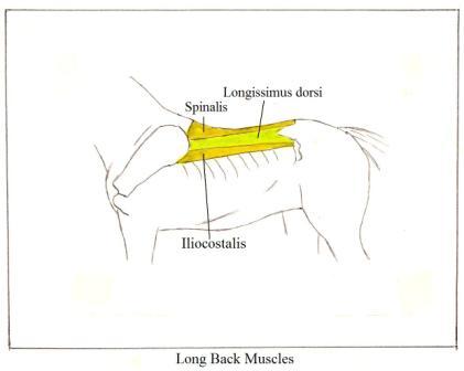
1. Long back muscles. Click
here for picture. These muscles are some of the most important
ones that you will deal with in massaging a horse. They are the major support and protection for
the spinal vertebrae and are subject to major problems involving tightness,
soreness and muscles spasms from multiple sources. They are also the extensors that have to
lengthen when the abdominal muscles contract.
Technically,
these muscles are part of the second layer, but the upper-most, superficial
layer that covers them is primarily a thin sheet of connective tissue.
There are three
of these muscles on either side of the spine.
Starting closest to the spine and moving outward, they are the Spinalis, Longissimus dorsi and the Iliocostalis. Their forward attachments are to the withers and
the ribs under the shoulder blade (scapula).
They run the entire length of the back and attach to the forward edge of
the pelvis.
The Iliocostalis is easy to feel where it attaches to the
pelvis because its attachment is on the point of the hip and its outer edge,
between the point of the hip and the last rib; it feels like a hard, ropey
ridge. Some equine anatomy books show
these as one single muscle, but there are really three. The human body has these same muscles,
positioned in the same way and their purpose is the same—support and protection
of the spine and to extend as the abdominal muscles contract.
Sides of the Torso
2. Latissimus dorsi. This is a large flat muscle, somewhat
triangular in shape, that connects the horse’s back to
the humerus in the lower portion of the shoulder. The
origin portion of this muscle, which attaches along the spine, is connective
tissue. This is why we can palpate the
long back muscles under it.
The middle
portion contains the muscle fibers. The
insertion portion (attaching to the humerus) is
tendon (no muscle fibers). This is a
good example of a large muscle narrowing down to a tendon in order to attach to
a small point.
This muscle
offers support and control to the movement of the shoulder in its forward/back
swing. As the shoulder swings forward,
this muscle extends; its contraction helps bring the humerus
backward. Because this muscle connects
the back to the front legs, over-stretching of the front legs can put excess
stress on a horse’s back, and, conversely, problems in the back can affect
shoulder swing.
If you look at
this muscle on the human anatomy, its origin and tendon attachments and its
function are much easier to see.
Visualize an arm swinging forward (perhaps a tennis or baseball swing—any
throwing motion).
3. External oblique. Click
here for picture. This
is a large, relatively thin muscle that covers much of the rib cage. It starts out narrow at the point of the hip
and spreads out over the ribs. The top
edge attaches to the individual ribs.
The bottom edge merges into a band of fascia (connective tissue) that
then merges into the abdominal muscles.
This fascia provides a direct link between the external oblique and the
abdominal muscles and gives a rider a way of initiating a contraction of the
abdominals. The External oblique is the
muscle that lengthens and shortens the sides of the horse as it moves. These
muscles, on both sides of the horse, are important for correct bending. The movement in the lower edge of this muscle
is easy to see when a horse is trotting.
These muscles also expand and contract in time with a horse’s breathing. In a horse that has heaves, the lower edge of
this muscle is very noticeable in its contraction; we call it the “heave line”.
4. Internal oblique. Click
here for picture. This muscle could be thought of as both a
muscle on the side of the horse and one in its lower torso area (belly
line). The loin/flank area of the horse
has no bony support other than the lumbar vertebrae; there are only muscles
supporting and holding up this portion of the abdomen (akin to out lower
abdomen). The major muscle in this area,
and one accessible for massage, is the Internal oblique. It starts out narrow at its attachment to the
point of the hip and then broadens out as it swings down to the mid-line of the
belly. It acts as a sling, holding up
the contents of the abdominal cavity. It
encompasses the area we call the flank and groin. Tightness and muscle spasms are common in
this area and it is an important area to work on, but it is also one of the
most difficult. Many horses, in response
to pressure here, will kick first and ask questions later. Work in this area should be approached with
care.
The arrangement of the Internal and
External oblique muscles in humans is somewhat different. In the horse they are quite distinct from
each other. In humans they are close
together and actually over-lay one another, each going in a different diagonal
direction. But their function is
essentially the same. They are important
for longitudinal and lateral flexion of the torso.
Lower Torso (belly muscles)
5. Rectus abdominis. The Rectus abdominis
(abs) are a line of muscles on either side of the center line of the
belly. They are connected to the lower
edge of the External oblique by fascia.
In both horses and humans the Rectus abdominis
are long, relatively narrow muscles that attach to the pubic bone of the
pelvis, run along both sides of the center line of the belly and then attach to
a number of the ribs. In the horse their
contraction exerts a pull on the pubic bone that draws the pelvis down as the
hind legs swing forward. Their
contraction also lifts and supports the long back muscles, supporting and
holding the torso up from underneath.
Good abdominal muscle tone is as important for the horse as it is for
humans in keeping the back healthy and working correctly. Our term for a horse with poor abdominal
strength is “hay belly”; for people the term is “pot belly”. Click
here for picture – chest and abdominal muscles.
2.
THE HIND QUARTERS WITH THE HIND LEGS
The muscles of the hindquarters that I’ll be describing are listed
below. Again, I’ll start at the top and
work down.
1. Gluteal muscles—Gluteus medius
and Gluteus maximus
2. Tensor fascia latae
3. Quadriceps—Rectus femoris,
Vastus medialis, Vastus Intermedius, and Vastus lateralis
4. Hamstrings—Bicepts
femoris, Semitendinosus, Semimembranosus
5. Adductors
6. Gaskin
7. Gastrocnemius and lower hamstring attachments
Click
here for picture of superficial haunch muscles.
Click
here for picture of deep haunch muscles.
All of the
muscles we will be talking about are present in both horses and humans. There are some important variations, but the
basic similarity in muscle structure would be very easy to see if the horse’s
hind legs were straightened out like human legs rather than being folded under
the pelvis. But this folding is one of
the things that give the horse’s hindquarters much of their power.
The muscles of
the haunches are bulky, dense, powerful muscles whose primary function is
propulsion. These are power muscles;
their job is to send the horse forward.
They are easy to massage because of their size and accessibility.
1. Gluteal muscles. There are three gluteal
muscles, but one, Gluteus minimus, is deep and not
accessible to us for massage. The other
two, Gluteus medius and Gluteus maximus,
form the bulk of the muscle mass on the top of the haunches. (Other names you might see for these muscles
are middle glute for Gluteus medius
and superficial glute for Gluteus maximus.) Together these two muscles attach to the edge
of the pelvic bone where it joins the loins (iliac crest) and along the edge of
the sacrum. They both converge and
attach to the upper portion of the femur at the hip joint. Their contraction and extension works to
stabilize the forward/backward swing of the horse’s hind legs. These muscles are arranged the same way in
the human body and they perform the same function.
2. Tensor fascia latae. This muscle is important in the working of
the stifle. It originates from the point
of the hip as a muscle and then becomes fascia (connective tissue) as it
approaches and covers the outside of the stifle. It helps to support the stifle. Its contraction, along with the contraction
of the quadriceps, pulls the stifle forward and its extension allows the stifle
to move backward. The best results from
massage on this muscle would be in its upper portion where there are muscle
fibers. Humans have this same muscle and
it has the same function as it does in the horse. It also originates on the point of our hip as
muscle and becomes a strong band of fascia (the iliotibial
band) that attaches to the tibia bone just below the knee joint. It supports the muscles that lie on the
outside of our leg as well as supporting the knee.
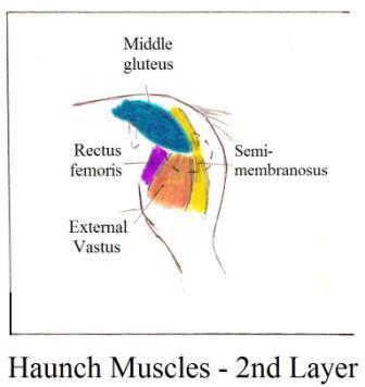
3. Quadriceps. In the horse these are four large muscles on
the front of the femur that are important for correct functioning of the
stifle. They are covered by the fascia
of the Tensor fascia latae, but they are still
accessible for massage. They are the
three vastus muscles (Vastus
medialis, intermedius, lateralis) and the Rectus femoris. They work
in conjunction with the Tensor fascia latae to bring
the stifle forward. The best access to
these muscles is an area just in front of the hip joint and on the inside of
the stifle joint. Humans have these same
muscles and they do the same job of bringing the human knee forward, but there
is one significant difference. In the
human, all four muscles converge into one large ligament that crosses the knee
and is known as the patellar ligament.
In the horse the attachments of these muscles at the stifle are separate
and distinct.
4. Hamstrings. If the quadriceps and the Tensor fascia latae pull the stifle forward, something has to pull it
back; the hamstrings do this. They are a
group of three muscles that run down the back of the horse’s haunch. Starting on the outside of the haunch and
moving around to the inside, they are the Biceps femoris,
Semitendinosus, and Semimembranosus. The Biceps femoris
is a large, fairly broad muscle with its origin on the sacrum. It crosses over the hip joint and swings to
the outside of the haunch for its lower attachment. The Semitendinosus
and Semimembranosus are long, but bulky muscles, also
with their origins on the sacrum, that go down the back of the haunch. Their lower attachments are in the area where
the haunch tapers into the hind leg.
When these muscles contract they pull the leg backward. The agonist/antagonist action
of these two muscle groups (quadriceps and hamstrings) produce the swing
of the hind legs in both horses and humans.
But there is a major difference in horse and human anatomy in the
arrangement and position of the hamstrings.
In humans the hamstrings have their upper attachment (origin) at what we
refer to as our “seat bones” (Ischial tuberosity). With
the attachment here, our legs swing from the hip joint without changing the
vertical position of our pelvis. In the
horse these muscles continue on past the Ischial tuberosity (the point of the buttock, or the horse’s seat
bones) and attach to the sacrum. With
the hamstrings positioned in this way, when they lengthen in response to the
contraction of the quadriceps, this extension goes all the way up to the
sacrum, not just to the point of the buttock.
Lengthening in this way allows the horse’s pelvis to be pulled downward
from its normal horizontal position and sets the horse’s hind legs up for a
powerful reach under the body and then a powerful push that sends the horse
forward. This dropping of the pelvis as
the hind legs come forward is what creates “engagement”. These three muscles are fully accessible and
respond well to massage over their entire surface.
The last three muscles, or muscle groups, of the hindquarters are easy
to find and massage.
5. Adductors. The adductors are the big muscles of the
haunch that are between the hind legs.
The biggest muscle, and the one you’ll make the most contact with, is Gracilis. Tightness
in these adductors can restrict the swing of the hind legs. These muscles are positioned the same in
humans.
6. Gaskin.
Horsemen use the term “gaskin” for two small muscles on the front of the
hind leg. The gaskin is actually two
small muscles whose tendons run all the way down the front of the hind leg to
the hoof. These muscles are important
because any tightness or soreness in them can exert tension on their tendons in
the lower leg. Release of tension in
these muscles can help relieve tension on the tendons.
7. Gastrocnemius.
The gastrocnemius is on the back of the hind
leg. It is also a small muscle with an
important tendon. You’ll find the muscle
belly just below the hamstring’s lower attachments. Its tendon attaches to the top of the hock. In humans this is known as the Achilles
tendon. Tension in this muscle can
stiffen hock movement.
3.
THE FOREHAND, SHOULDERS
This section of
the lesson on muscle anatomy is not intended to deal with all the muscles of
the horse. This is especially true of
the forehand because many of the muscles are not accessible to us for
massage. It is only the important
muscles that we can get to that I’ll be describing.
What we call
the forehand of the horse is actually the front portion of the rib cage (from
the withers and shoulders to the front of the chest), the shoulders and the
front legs.
Some of the
muscles of the forehand support the bottom of the rib cage, some are
responsible for moving the shoulder blades and front legs, and some hold the
shoulder blades in place against the ribs.
I’ll use these three divisions for my descriptions.
Support Muscles
There are four
pectoral muscles in the chest area and three of these are the major support for
the bottom of the rib cage. The Rectus abdominis muscle supports the torso from the pubic bone to
the beginning of the rib cage. The largest of the pectoral muscles takes over
support where the Rectus abdominis ends. It is a long, fairly large muscle that
attaches along the entire length of the breast bone (sternum) and then, close
to the elbow joint, it attaches to the humerus. When this muscle contracts,
it elevates the rib cage at the withers.
This muscle acts in much the same way as the human Pectoralis
major.
Looking at this muscle on a human anatomy
chart will give a clear view of how it also looks on the horse. Two other pectoral muscles also attach the
front legs to the body of the horse; they also go from the rib cage to the humerus. One can be
found directly between the front legs.
The other is in the front of the chest; what we would normally call the
chest muscle.
These muscles
are easy to find and massage.
Muscles That Move the Shoulder Blade
and Front Legs
I’m only
dealing with major muscles and I’m describing them as groups of muscles that do
a specific job.
The concept in
moving the front legs is simple—the muscles located in front of the shoulder
pull the humerus forward; muscles behind the shoulder
pull the humerus back. The major muscles involved in pulling the leg
forward (ones that we can access and massage) are the Biceps brachii and the lower portion of the Brachiocephalicus. The Biceps brachii
is in the front of the chest, next to the chest pectorals.
The Brachiocephalicus is a long muscles
that originates just behind the horse’s ear—on the first vertebrae (the
Atlas). It goes down the entire length
of the neck, following the contour of the neck vertebrae, then over the point
of the shoulder and inserts on the humerus. It is a prime mover in the process of
bringing the leg forward.
Massage on the
lower portion of the neck and lower portion of the shoulder will relieve
tension in the lower portion of this muscle as well as other important muscles
of this area.
The important
muscles that pull the leg back are the Latissimus dorsi and the Triceps.
I described the Latissimus dorsi
when describing the muscles along the back of the horse’s torso because of its
attachment, as fascia, along the thoracic vertebrae. Its muscle belly (the portion with muscles
fibers) is behind the shoulder and its strong tendon attaches to the humerus. Its
contraction exerts a strong backward pull on the humerus. Because the muscle fibers of this muscle are
behind the shoulder and just under the skin, massage to this area can be
beneficial in increasing shoulder mobility.
The triceps
pull the front leg back in a different way.
This muscle forms the back portion of the shoulder (the big, fleshy
portion behind the scapula) and is triangular in shape. Its origin attaches all along the back edge
of the scapula and also at two points on the humerus,
and then all the muscle fibers converge to attach at the point of the
elbow. When this muscle contracts it
closes the angle between the scapula and the humerus,
and thereby pulls the humerus backward.
Muscles That Hold the Scapula Against the Rib Cage
There are two
groups of muscles that keep the scapula in place against the rib cage.
One group is
between the rib cage and the scapula.
The other group is on the outside of the scapula. In both instances (inside or outside) the
process of stabilizing the scapula against the rib cage occurs at the top of
the scapula—leaving the bottom free to swing forward and back.
The important
muscles between the scapula and the rib cage are the Rhomboid and Serratus.
The Rhomboid
attachment to the scapula is on the inside upper edge. The other end of this muscle attaches to the
withers. So, the scapula is hanging from
the withers, and the Rhomboid is the attachment between the two. This muscle is the same in human
anatomy. Tightness of this muscle in the
horse can adversely affect the balance of the entire forehand. There is also a neck portion of the Rhomboid,
which I will discuss with the neck muscles.
Below the
Rhomboid attachment on the inside of the scapula is the attachment of the
Serratus. At its origin at the top of
the scapula it is a small, dense muscle mass that then divides into 13 long,
slender muscles. These 13 muscles attach
the top of the scapula to the middle of the rib cage on the first nine ribs and
to the last four neck vertebrae. This
arrangement of the Serratus is important because when the Serratus is working
correctly it causes the neck to stretch down and helps to bring the back
up. Much of this muscle arrangement is
cover by the scapula or heavy, dense muscles, but we have access to some portions
of these individual muscles that attach to ribs six through nine, and can also
make contact with those attaching to the lower neck vertebrae.
Because these
thirteen “fingers” of the Serratus all originate from a common muscle mass, we
can have a positive effect even on the sections we can’t reach if we can get
some of these “fingers” to release tension.
This is why relaxing the neck helps soften the rib cage and vice
versa. The human Serratus that is
accessible to massage only attaches the scapula to the ribs, it does not go
into the neck vertebrae.
Click
here to see the serratus with the shoulder blade in place.
Click
here to see the serratus with the shoulder blade removed.
Click
here to see the serratus in the extended state.
Click
here to see the serratus in the contracted state.
There is only
one major muscle on the outside of the scapula that holds it in place—this is
the Trapezius.
It stabilizes its top portion, while still allowing the bottom to swing
with the movement of the front legs. The
form and function of the Trapezius is the same in
humans and horses. It is a big
triangular muscle that originates along the spine of the scapula and fans out
in two directions to attach to the horse’s topline. One section attaches to the withers and the
area of the back just behind the withers.
The other section goes up the crest line of the neck covering most of
the Rhomboid muscle. This muscle is a
powerful anchor and stabilizer for the scapula, as well as support for the
neck.
The Trapezius is an extensor and when working correctly the
extension of the Trapezius allows the neck to arch
out in front of the horse’s torso. But
if the Trapezius contracts it will cause the neck to
arc upward, resulting in what horsemen call ewe-necked’ or “star-gazer”. This contraction of the Trapezius
will also pull the horse’s back down in the portion under the saddle. (Notice that the result of this contraction
is the opposite of what happens when the Serratus contracts and lifts the
back.)
There are two
other muscles of the shoulder that I want to mention because their purpose is
different than the others I’ve described and they are big enough that massage can
be beneficial even though they are covered by other muscles of the
shoulder. They are Supraspinatus
and Infraspinatus.
These are long
muscles that run lengthwise down the scapula, one on either side of the spine
of the scapula. They are attached to the
scapula along almost their entire length so that there is very little extension
possible except at their tendons. These
strong tendons cross the shoulder joint (point of the shoulder) and attach on
the humerus side of this joint. The purpose of these muscles and their
tendons is to support the shoulder joint in its proper position against the rib
cage.
As
you have probably gathered, there is a complexity of muscles contained in a
fairly small area in the shoulders, and often it can be difficult to isolate
exactly which muscles you may be massaging.
Fortunately, massaging the bulk of the shoulder and the areas behind it
and in front of it will benefit all of them.
The reason for
describing them in detail as I did was to give you a better understanding of
how the shoulders and front legs are organized in relationship to the rib cage
and how they work to produce movement.
4.
THE NECK
Neck
Comparing human
and horse anatomy in the neck will show basic similarities in muscles and structure,
but the extension of the neck hanging out in space in front of the horse
requires some special adaptations, especially in deep muscles and ligaments.
At the deep
level these systems of supporting ligaments and muscles are not accessible for
massage and, in the case of ligaments, do not readily respond to massage.
I’m only describing the superficial and
second levels that we can massage, but releasing tension in these superficial
muscles can gradually have an effect on the deeper muscles as well.
Muscles along
the crest are going to be support muscles, muscles that hold the neck up. There is a very strong ligament that forms
the crest line of the neck (the Nuchal ligament); it
runs from the withers to the poll, and it is to this ligament that the
supporting neck muscles attach.
The important
muscles attaching to this ligament are the cervical (neck) portions of the
Rhomboid and Trapezius and the Splenius. The Rhomboid and Trapezius
attach to the Nuchal ligament and connect the crest
line of the neck to both the rib cage and the scapula. The Splenius supports the side of the neck;
it starts at the withers and spreads out to attach to the upper neck
vertebrae. All of these muscles are easy
to massage. Click
here for picture.
Supporting the
vertebrae of the lower portion of the neck is the cervical portion of the
Serratus. (Remember that the Serratus
attaches to the ribs as well as to the neck vertebrae.) This cervical portion of the Serratus goes
from its origin on the under side of the scapula to the last four neck
vertebrae. This is a very important
muscle for obtaining correct carriage of the neck. Its attachments to the neck vertebrae occur
where the neck has its downward arch. If
these muscles have correct tension, their contraction will lift these lower
neck vertebrae, causing the crest line muscles to extend and allow the topline to lengthen.
If muscle tone
is poor in these cervical Serratus, it allows this portion of the neck to
“sag”, causing a dip in the crest line of the neck. Horsemen refer to this configuration as
“ewe-necked” or “star-gazer”. Correct
function of the Serratus has a significant effect on how the horse is able to
use the rest of its body.
The Serratus
attachments to the lower neck vertebrae are under the Brachiocephalicus,
but can be massaged through it.
The human
Serratus also divides up into separate muscle “fingers”, but they attach only
to the ribs, there are no neck attachments.
The Brachiocephalicus is a long muscle on the lower side of the
neck. It starts behind the horse’s ear,
follows the contour of the neck vertebrae, goes over the point of the shoulder
and finally attaches to the humerus. Its contraction is involved with the
forward/back swing of the front legs as well as giving support to the neck from
the bottom, much as the abdominals and pectorals support the torso.
There is one
other muscle that can cause tension in the neck. This is the fourth pectoral. You will remember that the other three
pectorals attached the humerus to the rib cage. This fourth pectoral lies lengthwise along
the front edge of the scapula and forms another attachment of the scapula to
the humerus.
It can be felt as a long, tight ridge in front of the scapula, and
tension in this muscle can easily prevent any softening and lengthening of the
neck.
Massage to all
of these neck muscles produces positive responses not only physically, but
mentally as well.
Head
Because of its
endorphin points, massage to the head, especially the ears and poll, can be a
powerful relaxing experience for the horse.
I have been told that cavalry officers would “pull” their horse’s ears
in-between cavalry charges to relax them.
This relaxing head massage can be especially beneficial for nervous
horses and is also a nice finishing touch to any kind of equine massage, except
pre-event stimulation massage.
The work I do
on the head is primarily to release tension in the jaw line and balance the jaw
joint and poll.
There
is a long, thin muscle (Zygomatic) that runs from the
corner of the horse’s mouth up into the ridge of the cheekbone. Pressure to this muscle where it meets the
big cheek muscle (Masseter) will produce a relaxation
of the horse’s mouth and jaw line. This
relaxation in the mouth helps when you are doing work on the jaw joint. The jaw joint of the horse is found in the
same place as you would find it on the human skull—just in front and slightly
down from the ear.
Working to
release the jaw joint doesn’t involve so much of massaging muscles, but more of
releasing the joint itself.
In
addition to the massage work that involves the jaw, there are numerous small
muscles around the poll—behind it, on the forehead and around the ears, and it
is important to loosen these if you are going to obtain good poll flexion. Poor poll flexion can stiffen a horse
throughout its entire body.
The
other muscles I address when there is tension in the poll are the ones of the
throatlatch. Even when the poll muscles
are capable of extending to give good poll flexion, tight muscles in the
throatlatch area can prevent this flexion.
I think of it as trying to flex the head when there’s a tennis ball in
the throat area.
Horses that
crib often have difficulties with poll flexion because of over-developed, tight
muscles in the throat area.
This brings us
to the end of this survey of skeletal anatomy and the major muscles important
for support and movement that are accessible for us to massage. Lesson three will cover the horse’s gaits and
movement patterns.
Assignment:
The aim of this assignment is to enable
a student to become so familiar with the horse’s muscular and skeletal anatomy
that they can put a hand any place on a horse’s body and know what muscle is
under their hand, what the shape of that muscle is, where the origin and
insertion attachments of the muscle are, what bones are under that muscles and
what the job of the muscle is – what movement of the bones does the muscle
activate.
Use your anatomy books as you do this
assignment.
I am not looking for a comparison of
the conformation and muscle density of the three horses I’ve asked you to look
at. I’m asking you to look at three
horses in order to give you a number of horses to practice on for this
assignment.
On three horses, find the bones of the
skeleton that we covered in the lesson.
If a bone is covered by substantial muscle mass, be able to visualize
where it is and its shape. Touch the
bones where ever possible. Follow the
progression of the bones, starting with the head and going back to the
tail. Also, with finger tips, trace leg
and foot bones – fronts and hinds.
On three horses, find muscles of the
horse’s body that we covered in the lesson.
Touch these muscles, tracing the outlines with your finger tips whenever
possible. Start at the head and work
back to the tail.
After your exploration of the horse’s
bones and muscles, write a report describing your thoughts, ideas and any new
awareness about what you are feeling – perhaps new things you did not know
about horses and their bodies.
Did you have any “ah ha” moments about
things that suddenly made sense to you that you may not have understood
before?
I would like any thoughts that occur to
you as you are turning the descriptions of the muscles and bones in the lesson
into a tactile journey of the horse’s body.
Send your report to: eleanorblazer@horsecoursesonline.com
The original
instructor, Betty Lindquist, retired